The key to a better biopsy experience isn’t the technique itself, but your active participation in the process.
- Understanding the « why » behind pain, delays, and potential errors transforms patient anxiety into empowered action.
- Proactive steps taken before and after the procedure have a significant impact on healing and cosmetic outcomes.
Recommendation: Use the specific questions in this guide to open a clear dialogue with your care team about pain management, result interpretation, and aftercare from the very beginning.
Hearing that you need a biopsy to investigate a suspicious lump is a moment that stops time. The clinical terms—core needle, excisional, pathology—can sound foreign and intimidating, and the waiting period that follows can be filled with anxiety. Your mind likely races with questions about the procedure itself, the potential for pain, and what the results might mean. It’s also natural to worry about the physical aftermath, especially the risk of a permanent scar or indentation that serves as a constant reminder of this stressful time. While many guides focus on the medical definitions, they often overlook the most critical element: your role in the journey.
The common advice to « trust your doctor » and « wait for the results » is well-intentioned but can leave you feeling powerless. This guide is different. As a nurse navigator, my role is to empower you with knowledge, turning you from a passive recipient of care into an active partner in your diagnosis. We will go beyond the basics to explore the nuances of the biopsy process. We’ll discuss how you can advocate for your comfort, understand why results sometimes take longer than expected, and what to do if you’re concerned about a « benign » result that doesn’t feel right. The goal is to demystify the experience and provide you with concrete tools to manage not just the physical procedure, but the mental and emotional impact as well.
This article provides a clear roadmap for your biopsy journey, from managing immediate concerns like pain to understanding the long-term implications for your health and well-being. The following sections will equip you with the information and questions you need at every step.
Summary: Needle or Excisional: Which Biopsy Technique Minimizes Scarring Risks?
- How To Manage Pain During A Core Needle Biopsy Without Sedation?
- Why Pathology Results Take Longer For Rare Tissue Types?
- The « Sampling Error » Trap: Can You Trust A Benign Result?
- Blood vs Tissue: Is Liquid Biopsy Ready To Replace The Needle?
- How To Care For The Biopsy Site To Avoid An Ugly Indentation?
- Disposable or Autoclaved: Which Instruments Are Safer For Minor Procedures?
- The « Incidentaloma » Trap That Leads To Unnecessary Biopsies
- Why A Multidisciplinary Team Increases Cancer Survival Rates?
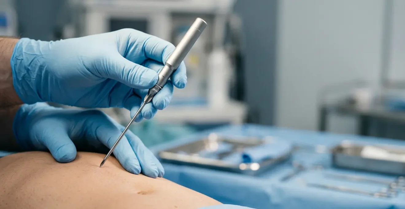
How To Manage Pain During A Core Needle Biopsy Without Sedation?
The fear of pain is one of the most immediate concerns when facing a biopsy. While core needle biopsies are typically done with a local anesthetic, « local » doesn’t always mean you’ll feel nothing. You may experience sensations of pressure, which are normal, but sharp pain is a signal that needs to be communicated. The key to managing this is not to passively endure it, but to have an informed dialogue with your radiologist or doctor before the first needle is even introduced. Your experience matters, and you are your own best advocate for your comfort.
Effective pain management begins with setting expectations. A good clinician will explain what they are doing at each step and what you should expect to feel. However, you can take an active role by asking specific questions beforehand. This establishes a partnership where you are encouraged to speak up. Remember, the goal of the local anesthetic is to make the procedure tolerable. If it’s not working effectively, the clinician has options, such as administering more anesthetic or adjusting their approach, but they can only act on information you provide. Feeling empowered to speak up is crucial for a better experience.
Your Action Plan: The Patient Empowerment Checklist for Pain Discussion
- Ask: ‘What is your protocol if the local anesthetic is not fully effective during the procedure?’
- Request: ‘Can we use a topical numbing agent before the injection to reduce initial discomfort?’
- Clarify: ‘What sensations should I expect – pressure versus sharp pain – and what should I report?’
- Discuss: ‘What pain management options are available in the hours after the local anesthetic wears off?’
- Confirm: ‘What are the red-flag symptoms (expanding hematoma, severe pain) that require immediate medical attention?’
Why Pathology Results Take Longer For Rare Tissue Types?
The waiting period between a biopsy and its results can be one of the most anxiety-inducing parts of the entire process. While many routine results come back in a few business days, it’s not uncommon for some to take much longer, stretching into weeks. This delay is rarely a sign of something being overlooked; rather, it often indicates the complexity and thoroughness of the diagnostic process, especially when dealing with rare or challenging tissue types. Understanding what happens to your tissue sample in the « black box » of the pathology lab can help demystify this wait.
Once collected, your tissue sample begins a meticulous journey. It is preserved, sliced into micro-thin sections, placed on glass slides, and stained with special dyes to highlight cellular structures. A pathologist then examines these slides under a microscope. For complex cases, this initial review might not be enough. The tissue may require additional, specialized stains to identify specific proteins or markers. If a rare condition is suspected, the sample might be sent to a sub-specialist pathologist with expertise in that specific area or require advanced molecular testing.
Case Study: The Reality of Advanced Molecular Testing
The American Cancer Society highlights a common scenario for extended delays. When tissue specimens require molecular tests like next-generation sequencing (NGS) at specialized central laboratories, results can take 2 to 3 weeks to return. This extended timeframe reflects the meticulous nature of advanced testing and the logistics of sending samples to expert facilities, ensuring the most accurate diagnosis possible.
This careful, multi-step process is designed to provide the most accurate diagnosis possible. So, while the wait is difficult, a longer turnaround time often reflects a commitment to diagnostic precision rather than a cause for alarm.
The « Sampling Error » Trap: Can You Trust A Benign Result?
Receiving a « benign » (non-cancerous) result after a biopsy is a moment of immense relief. But what happens when that result doesn’t align with what you’re feeling, or what the initial imaging suggested? This is a valid and important concern, rooted in a concept known as sampling error. A biopsy only analyzes the small piece of tissue that was removed. If the needle misses the most suspicious part of a lesion, the result could be a « false negative »—showing benign tissue when malignant cells are present nearby. While not common, it’s a possibility every patient should be aware of.
According to the Susan G. Komen Foundation, false negatives may happen in up to 4 percent of cases in image-guided core needle biopsies where the mass isn’t palpable. This is why a simple « benign » report isn’t always the end of the story. The most critical step is the conversation that happens *after* the result. When a pathologist’s report of benign tissue conflicts with a radiologist’s or clinician’s suspicion, it’s a red flag known as clinical-pathological discordance. This should automatically trigger a discussion about the next steps, which could include closer monitoring, a repeat biopsy, or an excisional biopsy to remove the entire lesion for a definitive diagnosis.
Your role in this is to ensure that conversation happens. Don’t be afraid to ask your doctor, « Does this benign result fully explain the imaging findings and my symptoms? » or « What is our plan for follow-up? » Trusting your body and advocating for clarity are essential parts of navigating your health. A benign result is wonderful news, but it should be a confident conclusion, not a lingering question mark.
Blood vs Tissue: Is Liquid Biopsy Ready To Replace The Needle?
The idea of a simple blood test replacing an invasive needle biopsy sounds like science fiction, but it’s a rapidly evolving reality in oncology. This technology, known as a liquid biopsy, works by detecting tiny fragments of cancer DNA (called circulating tumor DNA or ctDNA) that are shed from a tumor into the bloodstream. It holds incredible promise for the future of cancer care, but for now, it’s essential to understand its current role and limitations. It is not yet a replacement for the traditional tissue biopsy, but rather a powerful complementary tool.
As experts in the journal *Signal Transduction and Targeted Therapy* emphasize, the tissue biopsy remains the undisputed « gold standard. »
Tissue biopsy remains the gold standard for tumor diagnosis due to its high level of laboratory standardization, good consistency of results, relatively stable samples, and high accuracy of results.
– Ma, Liwei et al., Signal Transduction and Targeted Therapy
A tissue sample provides a wealth of information that a blood test cannot. It allows a pathologist to see the cancer cells in their environment, determine the tumor’s architecture, and assign a grade (how aggressive it looks), which are all critical for initial diagnosis and staging. A liquid biopsy, in contrast, cannot provide this level of detail. However, it excels in other areas, such as monitoring a patient’s response to treatment or detecting cancer recurrence, often earlier than imaging can.
The following table, based on information from the College of American Pathologists, helps clarify the distinct roles these two powerful diagnostic tools play in cancer care today. As you can see, their strengths are different and often complementary.
| Application | Liquid Biopsy Status | Tissue Biopsy Status |
|---|---|---|
| Initial Cancer Diagnosis | Not yet reliable – requires tissue confirmation | Gold standard |
| Monitoring Treatment Response | Excellent – serial sampling feasible | Limited – invasive to repeat |
| Detecting Recurrence | Excellent in known cancers | Challenging to repeat frequently |
| When Tissue Unavailable | FDA-approved for genotyping (EGFR, PIK3CA panels) | Not applicable |
| Tumor Grading & Staging | Cannot provide histologic detail | Essential for grading/staging |
| Low Tumor Burden Detection | Limited – requires sufficient tumor shedding | Direct sampling more reliable |
How To Care For The Biopsy Site To Avoid An Ugly Indentation?
While the primary focus of a biopsy is diagnosis, concern about the cosmetic outcome is completely valid. The fear of a permanent scar, divot, or indentation is a real source of anxiety. The good news is that you have a degree of control over the healing process. Excellent aftercare is crucial, but proactive healing begins even before your procedure. The steps you take to prepare your body can significantly influence tissue repair and minimize the risk of an undesirable cosmetic result.
Wound healing is a complex biological process that relies on good blood flow and adequate building blocks, like collagen. Several factors can either support or hinder this process. For instance, nicotine is a potent vasoconstrictor, meaning it dramatically reduces blood flow to the skin, which can starve the healing tissue of oxygen and nutrients. Similarly, proper nutrition is not just a vague wellness concept; it’s a critical component of wound repair. Vitamins and minerals like Vitamin C and Zinc are essential co-factors for collagen synthesis. By optimizing these factors beforehand, you are setting the stage for better, faster healing.
Here are some proactive steps you can discuss with your doctor to optimize your healing outcome:
- Optimize Nutrition: In the weeks leading up to your procedure, ensure you have an adequate intake of protein, Vitamin C, and Zinc to support collagen production.
- Review Your Supplements: Discuss discontinuing supplements that can increase bleeding, such as Vitamin E and fish oil, for about a week before the procedure.
- Avoid Nicotine: Ceasing all tobacco and nicotine products at least two weeks before and after the biopsy is one of the most impactful things you can do for wound healing.
- Discuss Suture Technique: For excisional biopsies, ask your doctor about using deep dermal sutures. These stitches support the underlying tissue and can help prevent the volume loss that leads to indentation.
- Plan for Rest: Arrange to limit strenuous activity, especially for biopsies on high-tension areas like the back or joints, to prevent the wound from stretching and the scar from widening.
Disposable or Autoclaved: Which Instruments Are Safer For Minor Procedures?
When you’re in a medical setting, it’s natural to trust that every instrument used is sterile and safe. But have you ever wondered about the difference between the shiny, reusable metal tools and the single-use disposable ones? For minor procedures like biopsies, the shift towards single-use, disposable instruments is a critical, and often invisible, layer of patient safety. This isn’t about cutting corners; it’s about eliminating risk and improving outcomes in ways that even the most rigorous sterilization cannot always guarantee.
The most obvious benefit is the absolute prevention of cross-contamination. While autoclaving (steam sterilization) is highly effective against bacteria and viruses, some infectious agents are notoriously resilient. As medical experts at StatPearls note, this is a lesson learned from the highest levels of surgery.
Prion diseases like Creutzfeldt-Jakob Disease have infectious proteins that can survive standard autoclaving, which has driven the universal shift towards disposable instruments for neurosurgery.
– StatPearls Medical Education, Skin Biopsy – NCBI Bookshelf
Beyond infection control, there’s another crucial benefit directly related to your healing and scarring: sharpness. A factory-new disposable scalpel or biopsy needle has a perfectly engineered, pristine cutting edge. This optimal sharpness allows for a cleaner cut with less trauma to the surrounding tissue. In contrast, reused instruments, even when perfectly sterile, can develop microscopic dulling or burrs over time. A cleaner cut means less tissue damage, which in turn leads to faster healing times and a better final cosmetic outcome. It’s a small detail that makes a significant difference in your recovery.
The « Incidentaloma » Trap That Leads To Unnecessary Biopsies
With the rise of high-resolution imaging like CT scans and MRIs, we are finding things we weren’t even looking for. An « incidentaloma » is exactly what it sounds like: an incidental finding, a mass or lesion discovered by chance during a scan for an unrelated issue. While finding something unexpected can be frightening, it’s important to know that the vast majority of these findings are benign. However, their discovery can set off a cascade of further testing, anxiety, and sometimes, unnecessary biopsies. This is the incidentaloma trap.
The challenge is distinguishing a harmless anomaly from something that requires intervention. This decision weighs the risk of the finding being malignant against the risks of the biopsy procedure itself, which include infection, bleeding, and the potential for sampling error. For many low-risk incidental findings, the safest and most appropriate course of action is not an immediate biopsy, but a strategy of « watchful waiting » or active surveillance. This involves monitoring the finding with periodic imaging to see if it changes over time.
If you’re faced with an incidental finding, it’s crucial to have a nuanced conversation with your doctor about risk. This is not about questioning their judgment, but about becoming an active participant in a decision that involves statistical probabilities and personal risk tolerance. Asking for data specific to your age and the finding’s characteristics, and explicitly comparing the risks of biopsy versus monitoring, can help you and your doctor make the most informed and appropriate decision, potentially avoiding a procedure you don’t need.
Key Takeaways
- Patient agency is key: Your active, informed participation can significantly improve your biopsy experience and outcome.
- Question everything respectfully: From pain management protocols to benign results that feel « off, » informed dialogue is your best tool.
- Focus on proactive healing: Steps taken before and after the procedure, such as nutrition and rest, are as important as the aftercare itself.
Why A Multidisciplinary Team Increases Cancer Survival Rates?
In the complex world of cancer diagnosis and treatment, no single specialist holds all the answers. The most effective, safest, and most advanced care comes from a multidisciplinary team (MDT), often called a tumor board. This is a collaborative meeting where experts from different fields—radiology, pathology, surgery, medical oncology, and radiation oncology—come together to review and discuss a patient’s case. This collective intelligence acts as a powerful safety net, ensuring every angle is considered and leading to better, more accurate diagnostic and treatment plans.
The impact of this collaborative review is profound. A diagnosis is not just a label; it’s a complex puzzle. As Dr. Charles M. LeVea of Roswell Park Comprehensive Cancer Center explains, pathologists themselves rely on this team approach for challenging cases. They present difficult specimens at consensus conferences to get other doctors’ opinions, ensuring the final diagnosis is as accurate as possible. This process of second, third, and fourth opinions is built directly into the MDT model.
The data confirms the life-saving value of this approach. At a leading institution like Roswell Park Comprehensive Cancer Center, it’s been shown that for cases reviewed from outside facilities, the diagnosis is changed or refined in 11 to 18 percent of cases after review by their multidisciplinary team. This statistic is not an indictment of other doctors; it’s a powerful testament to the value of collective expertise. It means that nearly one in five times, a team of experts looking at the same information together arrives at a more precise conclusion, which can fundamentally change a patient’s treatment and improve their chance of survival.
To ensure you receive the most accurate diagnosis and a comprehensive treatment plan, ask if your case will be reviewed by a multidisciplinary tumor board. It’s the standard of care at top cancer centers for a reason, and it’s a level of security every patient deserves.
Frequently Asked Questions About The Biopsy Process
What is clinical-pathological discordance and why does it matter?
Clinical-pathological discordance occurs when the biopsy result (pathology) shows ‘benign’ but the lesion appears suspicious to the doctor based on clinical examination or imaging. This red flag should trigger a discussion about re-biopsy or excision, as sampling error may have occurred.
What questions should I ask my doctor after receiving a benign biopsy result?
Ask: ‘Given the benign result, what is our monitoring plan?’ ‘What specific changes should I watch for that would prompt me to come back sooner?’ and ‘Does this result definitively explain all my symptoms?’
How does imaging guidance affect the reliability of a benign result?
A benign result is more trustworthy if the radiologist’s report confirms that the needle was accurately placed in the most suspicious part of the lesion using ultrasound, MRI, or CT guidance for deep biopsies.
What is the statistical likelihood of this incidental finding being malignant in someone my age?
Request specific data about malignancy rates for your age group and the characteristics of the finding. Many incidental findings have very low malignancy rates, especially in younger patients without risk factors.
What are the risks of the biopsy itself versus the risks of monitoring this for 6 months?
Ask for a comparison of potential complications from the biopsy procedure (infection, bleeding, sampling error) against the risk of a short-term delay. For low-risk findings, watchful waiting may be safer than immediate intervention.
Is there a non-invasive imaging test that could give us more information before proceeding to biopsy?
Inquire about advanced imaging options (high-resolution MRI, PET scan, contrast-enhanced ultrasound) that might better characterize the lesion and potentially avoid an unnecessary biopsy.